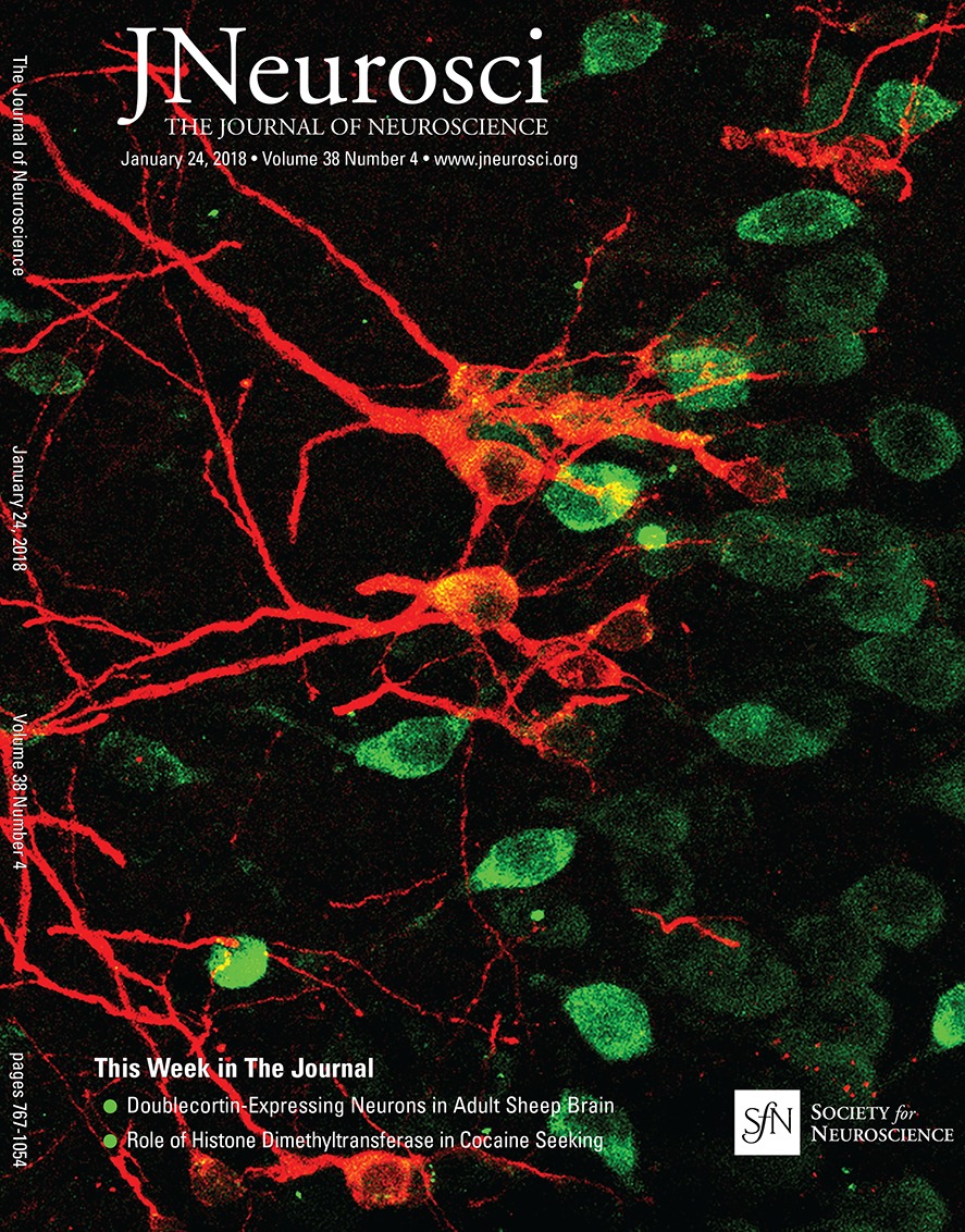
How Cells Fold the Cerebral Cortex
Víctor Borrell
Journal of Neuroscience 24 January 2018, 38 (4) 776-783; DOI:
How Cells Fold the Cerebral Cortex
Abstract
Folding of the cerebral cortex is as highly intriguing as poorly understood. At first sight, this may appear as simple tissue crumpling inside an excessively small cranium, but the process is clearly much more complex and developmentally predetermined. Whereas theoretical modeling supports a critical role for biomechanics,
experimental evidence demonstrates the fundamental role of specific progenitor cell types, cellular processes, and genetic programs on cortical folding.
Introduction
One of the most characteristic features of the human brain is its external folded appearance, due to the 3D arrangement of the cerebral cortex mantle forming outward folds and inward fissures. Folding patterns vary significantly between species, while being stereotyped within species. Developmental mechanisms responsible for cortical folding are an intriguing and elusive question that has remained poorly understood for decades. Initial hypotheses proposed that cortical folding relies on biomechanical forces,
but these can hardly account for the conservation of folding patterns within species nor their variability between species. A deep understanding of the cellular mechanisms of cortical development in the last 15 years has culminated in the identification of germinal layers, progenitor cell types, and developmental mechanisms specifically linked to cortical folding. This learning process has greatly benefitted from the ferret, a small carnivore emerging as a key model system to understand cortical folding. In this article, I review evidence and arguments supporting a central role for genetic and cell-biological mechanisms on the formation of cortical folds during development.
Comparative and experimental findings from a variety of species demonstrate that cortical folding depends critically on basal progenitor cells in the outer subventricular zone, particularly basal radial glia cells. Recent studies in ferret demonstrate that highly dynamic variations in gene expression levels within germinal layers, both during developmental time and across cortical space, have a fundamental impact in defining the prospective patterns of cortical folding. In order for me to review the cellular mechanisms responsible for cortical folding, I will first begin by defining what is cortical folding, and what is not.
Cortical folding
What is cortical folding?
Cortical folding is the developmental and histogenetic process that leads to a 3D conformation of the cerebral cortex mantle in a characteristic wrinkled external appearance, with rounded crests of cortical tissue separated by steep valleys, and a smooth internal surface.
Cortical folding is only found in mammals, and species with a folded cortex are named gyrencephalic. In contrast, lissencephalic species have a completely smooth cerebral cortex, lacking any kind of crest or valley with the only exception of the rhinal fissure, a brain landmark conserved across all mammals that separates the neocortex from the evolutionarily older pyriform cortex. As a general rule, cortical folding is naturally found in species with a large brain, such as cattle, monkeys, humans, elephants, and whales, whereas lissencephaly is typical of species with a small brain, such as rats or mice. Nevertheless, this simplistic generalization is not valid for a broad range of intermediate brain sizes, as discussed below.
The adult cerebral cortex is composed of six main neuronal layers (gray matter) orderly arranged from outside (layer 1) to inside (layer 6), plus the underlying white matter containing the fascicles of projecting axons. A cross section through a lissencephalic brain reveals that all cortical layers, including white matter, are smooth and roughly of constant thickness across their entire extent. In contrast, in gyrencephalic brains, the cortical layers are folded, conferring them the typical undulated appearance. All six neuronal layers are bent in each and every fold, and their thickness varies significantly between folds (thicker) and fissures (thinner). Importantly, the white matter is folded only at its border with layer 6, whereas its inner surface facing the ventricular cavity is smooth (
Fig. 1A);
hence, white matter is thicker at folds than at fissures. This is coherent with the fact that the number of neurons forming gyri is larger than those forming sulci, and so the number of axons descending down into the white matter is proportionally larger in gyri, leading to the mentioned thickening of white matter at folds. [..]
https://www.jneurosci.org/content/jneuro/38/4/776/F1.large.jpg?width=800&height=600&carousel=1
Pathological alterations of cortical folding documented in human patients as well as variations of cortical folding parameters observed across phylogeny (abundance, depth, or periodicity of folds) do not alter the smooth inner surface of the white matter, but only the outer (pial) surface of the cortex. Indeed, alterations of the ventricular surface, as in periventricular nodular heterotopia, seem to be mechanistically independent from cortical folding defects, as they are frequently observed in the absence of cortical folding alterations (
Barkovich et al., 2012). Intriguingly, some case studies have been reported where excessive gyration of the temporal lobe co-occurred with folding of the ventricular surface, forming diverticuli (
Hori et al., 1983), and ventricular expansion in the temporal and occipital lobes has been observed in the normal human brain. However, whereas ventricular folding may occur in the temporal and occipital lobes of the human brain, this seems not to be the case for all other cortical regions, nor it has been observed in other gyrated species analyzed; thus, it may not be a major mechanism driving gyrogenesis (
Sun and Hevner, 2014).
What is not cortical folding?
Multiple attempts have been made to model cortical folding in mouse because of its unmatched advantages as experimental animal. Most of these attempts have produced phenotypes that, while interesting, were far from bona fide gyrencephaly. Folding in these cases involves, for example, the overexpansion of the early cortical neuroepithelium with very limited neurogenesis (
Siegenthaler et al., 2009). As a result, this oversized monolayer of germinal cells folds onto itself affecting both the ventricular and the pial surfaces (
Chenn and Walsh, 2002;
Kingsbury et al., 2003), never observed in gyrencephalic species (
Fig. 1B). Other examples of mouse brain overgrowth that emulate cortical folding were achieved by blocking embryonic programmed cell death accompanied by exencephaly (
Haydar et al., 1999;
Depaepe et al., 2005), again unrelated to events occurring in natural gyrencephaly (
Reillo et al., 2011). Human cerebral organoids have also begun to be used to model and study cortical folding. Unfortunately, and similar to the early mouse models, folding of cerebral organoids has only been achieved so far by overexpansion and folding of the entire neuroepithelium, apical and basal sides, for example, by the overactivation of the mammalian target of rapamycin pathway (
Li et al., 2017).
These initial attempts were seminal because they
demonstrated experimentally that genetic mechanisms acting on progenitor cells in the developing embryo cortex could significantly modify cortical size and induce some forms of folding. This contributed to contradict a historical view that cortical folding is simply the inevitable result of the physical constraint of a limited cranial volume onto a growing brain (
Welker, 1990).
Unfortunately, these mouse models fail to properly reproduce natural folding as observed in gyrencephalic species. Conversely, other genetic manipulations in mouse lead to cortical overgrowth by increasing neurogenesis, and these also do not translate into cortical folding (Nonaka-Kinoshita et al., 2013). Importantly, also, these examples fail to fit into the biomechanical model currently accepted in the field, and substrate of further work: the differential expansion of an outer layer of the cerebral cortex over its inner core during development (see accompanying paper by Bayly and Kroenke).
Experimental models of bona fide cortical folding
Aware of the early failures to model cortical folding in the lissencephalic mouse, research has turned to use naturally gyrencephalic species to address this question at the mechanistic level. Work in human and nonhuman primate embryos has attracted much attention for its obvious relevance to understand human cortical folding (
Lui et al., 2011). It is most relevant to highlight work in the macaque monkey identifying new germinal layers (
Smart et al., 2002), several new types of cortical progenitor cells (
Betizeau et al., 2013), and
complex gene regulatory networks (Arcila et al., 2014), which have unraveled an outstanding complexity of cortical developmental mechanisms in primates (
Dehay et al., 2015). Some work has also been done in sheep, with particular relevance in clinical research (
Encinas et al., 2011). But the greatest advance in understanding mechanisms of cortical folding has come from studying the ferret. The unmatched advantages of this small carnivore over other species are several, including that it only develops primary folds in the cerebral cortex, that these follow a simple and highly reproducible pattern (nearly identical between individuals), that cortical neurogenesis proceeds after birth, and that cortical folds begin to form at 6–8 d of postnatal life. Hence, analysis and manipulation of naturally occurring cortical folds are much easier in ferret than in other experimental animals (
Fietz et al., 2010;
Xu et al., 2010;
Reillo et al., 2011;
Reillo and Borrell, 2012;
Nonaka-Kinoshita et al., 2013;
Kou et al., 2015;
de Juan Romero et al., 2015;
Masuda et al., 2015;
Martínez-Martínez et al., 2016;
Matsumoto et al., 2017).
Importantly, in the last 5 years, upon the emergence of new animal models for the development of cortical folds (
Reillo et al., 2011;
Borrell and Reillo, 2012), several new mouse models have also emerged that quite faithfully recapitulate key features of physiological gyrencephaly, namely, folding of all neuronal layers without folding of the ventricular surface. The first models relied on the acute and local manipulation of gene function by
in utero electroporation (
Stahl et al., 2013;
Florio et al., 2017), or gene overexpression by transgenesis (
Wang et al., 2016), and produced only one or two folds. Interestingly, a more recent study demonstrates the systematic and widespread folding of the cortex in a knock-out mouse line (
Del Toro et al., 2017), providing the first genetic mouse model for in-depth dissection of developmental mechanisms of cortical folding.
Developmental features linked to cortical folding
Folding of the cerebral cortex is linked to a number of developmental events, providing clues about potential mechanisms underlying this process. As mentioned above, cortical folding is consistently found in big-brained animals and not in small-brained animals, highlighting the importance of abundant neurogenesis and gliogenesis for folding (
Welker, 1990). Second, gyrencephaly is linked to splitting of the subventricular zone (SVZ) and the formation of an outer subventricular zone (OSVZ) during neurogenesis (
Reillo et al., 2011). The OSVZ is defined as a neurogenic germinal layer basal to the ventricular zone (VZ), and also basal to the inner subventricular zone (ISVZ), with unique cytoarchitectonics distinct from the latter (
Smart et al., 2002). However, some species deviate from this rule, such as the marmoset monkey or even rat, which are lissencephalic despite displaying a rudimentary OSVZ (
Kelava et al., 2012;
Martínez-Cerdeño et al., 2012). But even in such cases, the relative abundance of progenitor cells found in the OSVZ of a given species has a strong positive correlation with its Gyrencephaly Index (
Reillo et al., 2011;
Pilz et al., 2013), which is a measure of cortical folding independent from cortical size.
Thus, the relevance of OSVZ progenitors goes much beyond increasing neurogenesis to make a bigger brain, but to drive cortical folding. One important exception in this case is the macaque visual cortex, where doubled abundance of OSVZ progenitors doubles the amount and density of upper-layer neurons in A17 compared with A18, without increased folding (
Lukaszewicz et al., 2005).
Third, cortical folding is linked to a complex morphological and transcriptional diversity of basal progenitor cells in ISVZ and OSVZ, as opposed to those in the SVZ of rodents (
Noctor et al., 2004;
Englund et al., 2005;
Fietz et al., 2010;
Hansen et al., 2010;
Wang et al., 2011;
Reillo and Borrell, 2012;
Betizeau et al., 2013;
de Juan Romero et al., 2015;
Johnson et al., 2015). Fourth, folding of the cerebral cortex is linked to a high relative abundance of basal radial glia cells (bRGCs), as determined by comparing between mouse, marmoset, ferret, and sheep (
Wang et al., 2011;
Pilz et al., 2013). bRGCs were first identified independently by three laboratories (
Fietz et al., 2010;
Hansen et al., 2010;
Reillo et al., 2011), and also named intermediate radial glia cells (
Reillo et al., 2011) or OSVZ radial glia-like cells (
Hansen et al., 2010), more recently referred to as “outer radial glia” cells (
Gertz et al., 2014). Finally, the formation of primary folds in ferrets and humans is linked to regional differences in density of progenitor cell proliferation (especially basal progenitors) (
Reillo et al., 2011) and gene expression levels (
Stahl et al., 2013;
de Juan Romero et al., 2015;
Del Toro et al., 2017).
Intriguingly, there are few, but remarkable, exceptions to these general principles, which, rather than invalidating them, highlight the complexity of the cortical folding process. For example, regarding brain size, human patients with microcephaly may develop a reduced number of cortical folds with a normal appearance (depth, periodicity), even though brain (and cortex) size in these patients is smaller than average; and vice versa with lissencephaly (
Hong et al., 2000;
Baala et al., 2007;
Borrell and Reillo, 2012;
Bahi-Buisson et al., 2013;
Gilmore and Walsh, 2013). Similarly, species with similar brain volumes may be completely lissencephalic or gyrencephalic (i.e., manatee and chimpanzee, capybara and beaver, weasel and muskrat, ferret and marmoset) (
Welker, 1990;
Pilz et al., 2013).
It is fundamental to highlight that all these developmental features typical of gyrencephalic species may precede actual cortical folding by a long time, observable in the embryonic cortex, even only while it is still smooth.
Just like any other developmental process, cortical folding should not be analyzed merely under the perspective of what is going on while it occurs (Neal et al., 2007), but as the result of a series of events occurring along developmental time with cumulative effects. For example, whereas the actual folding of the cortical tissue occurs quite late in development (usually after neurogenesis and neuronal migration) (
Smart and McSherry, 1986;
Neal et al., 2007), it requires large numbers of neurons, which requires abundant cell proliferation and neurogenesis, which is determined by gene expression (
Fernández et al., 2016).
Thus, considering the entire temporal dimension of cortical development is fundamental to identify all the causative elements of cortical folding and to understand the specific contribution of each of them.
Developmental events causative of cortical folding (or smoothing)
While cortical folding was soon identified as the result of a developmental process, early attempts to identify causative events were driven by a strong intuitive notion: that the limited cranial volume may exert mechanical compression on the growing brain, and this drive tissue folding.
However, experimental evidence demonstrates that alleviation of cranial pressure does not lead to loss of folds, frequently rather the opposite (Welker, 1990).
Recent research has primarily focused on testing the role of progenitor cell proliferation, both in naturally gyrated and in naturally lissencephalic species. Recent studies in ferret demonstrate that the OSVZ emerges from the VZ only during a very brief period of embryonic development, after which this OSVZ must self-amplify for further expansion without additional contribution from VZ or ISVZ. Blockade of proliferation precisely during this brief critical period minimizes OSVZ formation, and it has a profound impact reducing cortical folding (
Poluch and Juliano, 2015;
Martínez-Martínez et al., 2016) (
Fig. 2A,
B). Similarly, the specific manipulation of OSVZ proliferation in ferret demonstrates its key role in cortical folding. Partial reduction in the mitotic activity of OSVZ progenitor cells reduces the size of cortical folds (
Reillo et al., 2011),
and augmented proliferation increases the number of folds and gyrencephaly index (Nonaka-Kinoshita et al., 2013; Masuda et al., 2015) (
Fig. 2C). It should be noted that some of the manipulations used to reduce OSVZ proliferation and cortical folding involved the perturbation of thalamic projections to the developing cortex (
Reillo et al., 2011). Whereas this manipulation may cause other, yet unidentified, alterations in cortical development that could lead to reduction of cortical folds, the complimentary gain-of-function experiments performed by
Nonaka-Kinoshita et al. (2013) argue that reduction in OSVZ proliferation may be a leading cause in the reduction of cortical folds also in this case. Together, these results demonstrate the central relevance of basal progenitor cells in OSVZ in the process of cortical folding.
Importantly, enhancing proliferation of basal progenitors in the mouse embryonic cortex is not sufficient to induce cortical folding (Nonaka-Kinoshita et al., 2013), which demonstrates the necessary involvement of other factors that distinguish between the development of gyrencephaly and lissencephaly. [..]
https://www.jneurosci.org/content/jneuro/38/4/776/F2.large.jpg?width=800&height=600&carousel=1
One essential difference in cortical development between mouse and ferret is the composition of basal progenitor cell populations. In gyrencephalic species (e.g., human, macaque, and ferret), the OSVZ and ISVZ are largely populated by bRGCs (Pax6+), whereas Intermediate Progenitor Cells (IPCs) (Tbr2+/Pax6−) are a minority (
Fietz et al., 2010;
Reillo and Borrell, 2012;
Betizeau et al., 2013;
Martínez-Martínez et al., 2016). This difference has contributed to the notion that a high proportion of bRGCs may be mechanistically important for cortical folding (
Reillo et al., 2011;
Borrell and Reillo, 2012;
Borrell and Götz, 2014). Accordingly, increasing the proportion of bRGCs in the developing mouse is sufficient to induce cortical folding (
Fig. 2).
Specific increase in bRGC abundance in the mouse embryo cortex has been achieved by using different genetic manipulations: loss of function of the nuclear protein Trnp1 (Stahl et al., 2013), activation of Shh signaling (Wang et al., 2016), and overexpression of the hominoid-specific genes ARHGAP11B (Florio et al., 2015) and TBC1D3 (Ju et al., 2016; Florio et al., 2017). All of these manipulations result in the formation of folds in the mouse cortex. In summary, increased neurogenesis is insufficient to drive cortical folding, but this is achieved by increasing the relative abundance of bRGCs, which profoundly alters the radial fiber scaffold (
Borrell and Reillo, 2012). Alternative mechanisms of cortical folding, solely relying on IPCs and independent from bRGCs, have also been proposed, but direct evidence is so far limited (
Rash et al., 2013).
Cortical folding has two components: the formation of folds and of fissures. The findings mentioned above demonstrate cellular correlates and mechanisms driving local cortical expansion, which result in the formation of outward folds.
A recent study proposes the existence of a complementary mechanism, driving the formation of fissures (Del Toro et al., 2017). An imbalance in the adhesion-repulsion forces between newborn neurons, as they migrate to the cortical surface, results in interspersed and selective cell clustering with increased migration speed, ultimately establishing the formation of cortical fissures. This study demonstrates that this mechanism drives cortical fissuring in the mouse cortex, and also that the same molecular mechanism may participate in folding of the ferret and human cortex (
Del Toro et al., 2017).
Importantly, these findings demonstrate that cortical folding involves not only outward growth, as initially proposed by Smart and McSherry (1986) and later supported by us and others (Borrell and Götz, 2014), but also the coordinated action of inward fissuring mechanisms.
Order versus chaos: conservation of folding patterns
Early studies of brain folding, and many of the mathematical models, care mostly about the fact that the cortical tissue folds but largely ignore the exact pattern in which it folds (
Toro and Burnod, 2005;
Xu et al., 2010;
Tallinen et al., 2014;
Mota and Herculano-Houzel, 2015).
In contrast, mounting evidence demonstrates that folding patterns are very similar between individuals within a species, and quite remarkably conserved between species within a clade. Although this is clear for species with simple folding patterns, such as ferret or cat (
Borrell and Reillo, 2012), at first sight it does not strike as being the case for species with a highly convoluted cortex like humans.
Similar to the branches of a tree, in highly folded brains, cortical fissures follow a hierarchical organization: primary fissures are the deepest and form first during development; later these are subdivided by secondary fissures; then these by tertiary fissures, and so on. In humans, the sulcal pits of primary fissures (their deepest point) have nearly identical location across individuals (
Lohmann et al., 2008). Smaller animals with simpler patterns (e.g., ferret) only develop primary fissures, and their pattern is robustly conserved across individuals (
Welker, 1990). These remarkable facts strongly suggest the influence of a deeply imprinted deterministic program, possibly genetic.
The location of secondary fissures is less well conserved, and those of yet higher order appear to be randomly formed, suggesting a limited (or absent) genetic/deterministic influence on those. Strikingly, the similarity of folding patterns between monozygotic twins (genetically identical) is statistically above the general population, including higher-order fissures (Lohmann et al., 1999). Together, these observations support the existence of genetic mechanisms regulating cortical development that robustly define the patterns of cortical folding, especially the first and deepest fissures while less robustly those of higher order. The increasingly greater variability among higher-order folds and fissures suggests that other factors may gain relevance in their patterning, particularly factors with a greater degree of stochasticity.
Mechanisms that define folding patterns
Once we understand that primary folding of the cerebral cortex is not random, but has an important component of hardwiring, it is time to ask what are the mechanisms that determine specific folding patterns. Current evidence points to two types of factors being involved: spatial and temporal.
Spatial factors
As I have discussed above, cortical folding is linked to, and depends on, the proliferation of cortical progenitor cells, particularly those in the OSVZ. Detailed analyses of progenitor proliferation in the developing ferret cortex, before the formation of folds, show the existence of regional variations in the density of cell proliferation and mitosis. This pattern of cell proliferation density correlates faithfully with the eventual location of primary folds (the only type found in ferret), with regions prospective of gyrus accumulating greater proliferation than regions prospective of sulcus (
Reillo et al., 2011). Similar correlations have been shown in species with more intricate cortical folding, such as cat and human (
Reillo et al., 2011), as suggested previously on hypothetical grounds (
Kriegstein et al., 2006). In these cases, the high density of OSVZ proliferation correlates with the expansion and folding of major cortical areas or lobes (i.e., parietal and temporal lobes vs cingulate cortex and insular cortex/sylvian fissure), which again is consistent with the patterning of primary folds and fissures.
To identify the possible genetic mechanisms regulating this patterned cell proliferation, a transcriptomics analysis was conducted in the developing ferret comparing germinal layers between prospective gyrus and sulcus (
de Juan Romero et al., 2015). From the thousands of genes found differentially expressed between these two regions, many are expressed in quite distinct modular patterns, where one region of high expression level is abruptly followed by an adjacent region of low expression level.
These modules of gene expression are prominently found in the OSVZ and faithfully map the prospective location of primary folds and fissures, strongly supporting their having a role in determining the pattern of primary cortical folds (de Juan Romero et al., 2015). Importantly, none of those genes is expressed in modules in the smooth mouse cortex during development, but they do in the embryonic human cortex. Indeed, genes driving folding of the mouse cortex when blocked (e.g.,
Trnp1,
Flrt1, and
Flrt3) are among those with modular expression patterns, with regions of low expression leading to gyrus, and vice versa (
Stahl et al., 2013;
Del Toro et al., 2017).
Finally, many of the modular genes are mutated in human malformations of cortical development, together supporting a key relevance of these genes and expression modules in defining cortical folding patterns (de Juan Romero et al., 2015).
Mechanisms driving the formation and maintenance of these modules of gene expression have not yet been identified, but one could argue that some or all of the known mechanisms of gene expression regulation may be involved in this process, including
chromatin remodeling, DNA modifications, histone acetylation and/or methylation, noncoding RNAs, and even direct regulation of mRNA stability. Very significant is the question of how these modules may arise in the first place. One interesting possibility on this regard is that the establishment of distinct borders of gene expression may occur following mechanisms similar to the establishment of stripes in the early
Drosophila embryo and progenitor cell subtypes in the mouse embryo spinal cord, including the existence of counter-gradients of transcription factors with mutually repressive activities (
Stanojevic et al., 1991;
Jessell, 2000).
Temporal factors
Time is also a critical factor in the development of cortical folds. Folding of the ferret cortex is abrogated by transiently blocking progenitor cell proliferation during a brief time window at mid-gestation (
Poluch and Juliano, 2015).
It has been recently shown that this 2 d time window (embryonic days 34–36 in ferret, out of 42 embryonic days) is a critical period for the initial seeding of the OSVZ with bRGCs generated in the VZ, which act as cell founders of this important layer (Martínez-Martínez et al., 2016). Blocking VZ proliferation or bRGC production during this critical period severely impairs OSVZ formation, which plays a fundamental role in cortical folding as discussed above.
In a similar way as spatial patterns of gene expression define the protomap of primary cortical folding (
de Juan Romero et al., 2015),
temporal variations of gene expression in VZ define the critical period for formation of bRGCs destined to initiate the OSVZ (Martínez-Martínez et al., 2016). Different sets of genes bookmark the opening and the closing of this critical period, so the prediction is that varying the timing of their expression would affect the duration of the critical period. In turn, this would profoundly alter the amount of founder progenitor cells and, consequently, the eventual size of the OSVZ and of cortical folds.
The role of biomechanics
Although I have highlighted in this review the relevance of cell-biological processes and their genetic regulation on cortical folding, it is quite clear that some aspects of this process escape cell biology and genetics.
This is best illustrated with the observation that the pattern of cortical folds and fissures in monozygotic twins, who are genetically identical, is not identical (Lohmann et al., 1999). Indeed, most of the arguments presented above regarding the importance of cell biology and genetics are only fully applicable to primary fissures. For example, variations in cell proliferation observed along the germinal layers of the human embryo cannot possibly account for the highly elaborate patterns of cortical folds in the adult human brain (
Reillo et al., 2011). Biomechanical forces acting on cortical folding are still poorly characterized and understood, but one possibility is that they may build upon the basic histological heterogeneity framework brought out by genetics and cell biology.
Local hydraulic pressures, tension forces, and such (Van Essen, 1997; Hilgetag and Barbas, 2006; Xu et al., 2010) may sculpt cortical topology on top of the basic pattern laid out by cell biology at earlier stages. Other processes occurring late in cortical development when folds grow and begin to acquire their final shape, such as gliogenesis and growth of neuropile, may also contribute to the shaping of cortical folding, whereas myelination has been suggested to have a limited effect (
Neal et al., 2007).
One intriguing feature of cortical folding is that the spatial periodicity of the smallest (highest order) folds is remarkably homogeneous along the cortex within any given species (and even between some groups of species), suggesting the existence of purely physical constraints to tissue buckling.
One of these may be cortical thickness, where a thicker cortex with more cellular elements stacked onto each other requires a wider bending angle than thinner cortices, as previously postulated (Zilles et al., 2013). This notion has been recently modeled (
Mota and Herculano-Houzel, 2015); but far from being generally accepted, it remains quite controversial (
Fernández et al., 2016;
Lewitus et al., 2016).
In conclusion, mechanisms underlying cortical folding have remained enigmatic and elusive since the discovery of this fascinating feature (
Welker, 1990). The advent of acute genetic manipulation and, most importantly, the use of appropriate animal models, such as the ferret, have recently provided the first empirical demonstrations of specific cellular mechanisms driving cortical folding (
Fernández et al., 2016). We should expect an exponential growth of knowledge on this field as technology progresses further, most especially on genome engineering and
in vitro fertilization of “nongenetic” species to produce mutant strains (
Kou et al., 2015). Using mouse as a model system has led to paramount advances in understanding the genetics and cell biology of human development, function, and disease. From now on, using new, more sophisticated and more appropriate model systems, such as ferret
in vivo, and possibly cerebral organoids
in vitro, will likely pave the way to understand some of the more complex features of the human brain, especially the development and folding of the cerebral cortex, and its complex malformations in developmental disease (
Barkovich et al., 2012;
Lancaster et al., 2013).


 www.sott.net

 cassiopaea.org


") .
.")