You are using an out of date browser. It may not display this or other websites correctly.
You should upgrade or use an alternative browser.
You should upgrade or use an alternative browser.
Mass Extinctions, Evolutionary Leaps, and the Virus-Information Connection
- Thread starter Pierre
- Start date
Chapter 11: Viruses Contribute to Mass Extinctions
Cometary events cause mass extinctions during which some species are eradicated while other species are spared and suddenly evolve.
As described in the previous chapter[1], viruses have species-specific destructive action, but there’s a difference between a virus causing diseases in a given species and a virus decisively contributing to the extinction of this same species.
So in the present chapter, we will attempt to answer the following question: Are there documented cases of pathogens in general and viruses in particular being notably involved in the extinction of a given species?
1/ Past virus-induced extinction
Let’s start with the previously described[2] K/T extinction ca. 66 Mya, the most recent and the most documented of the big five mass extinctions.
Infectious diseases are considered as one of the major cause of the elimination of some taxa during the K/T extinction while other taxa were being spared. These species-specific pathogens are fungi[3] [4], bacterias[5] and of course viruses[6].
The K/T extinction seems to have involved viral diseases which contributed to the mass extinction:
For example during the K/T extinction, the die off was not instantaneous but followed a pattern suggestive of spreading contagion. In fact, basing their findings on pathogens found within amber-entombed insects, Poinard has concluded that the extinction of the dinosaurs was due to "the cumulative, cascading effects of many diseases.
Poinard gave more details about how pathogens eradicated the dinosaurs along with numerous other Cretaceous species:
insect-vectored fungal and viral diseases were critical in determining which plants lived and died in the Cretaceous world.[7]
According to Poinar, insect-borne viruses, infected plants that, in turn, infected the dinosaurs:
Since extant aphids carry approximately 50% of insect-transmitted plant viruses, perhaps they were responsible for the rapid turnover of Late Cretaceous plant lineages.[8]
The K/T event is not the only mass extinction marked by the destructive activity of viruses. The cometary event that triggered the Younger Dryas 12,900 years ago, eradicated most of the woolly mammoths[9], but left pockets of survivors in Wrangel Island[10] who survived until ca. 4,200 BP[11]

© CC BY-SA 3.0
Location of Wrangel Island
The DNA of the Wrangel Island mammoths revealed some peculiarities:
Here, we show that a Wrangel Island mammoth genome had many putative deleterious mutations that are predicted to cause diverse behavioral and developmental defects. Resurrection and functional characterization of several genes from the Wrangel Island mammoth carrying putatively deleterious substitutions identified both loss and gain of function mutations in genes […] These data suggest that at least one Wrangel Island mammoth may have suffered adverse consequences from reduced population size and isolation.[12]
Now, the mutations found in the mammoths of Wrangel Island are attributed to the scarce population and the subsequent inbreeding. However inbreeding doesn’t cause genetic defects per se but only increase the risk of recessive gene disorders[13].
More importantly, the wooly mammoths from Wrangel Island revealed gain of function mutations, which is difficult to explain by an inbreeding process, but, as shown later[14], can easily be induced by viruses.
According to the senior curator of the American Museum of Natural History, Ross MacPhee it is indeed a viral epidemics[15] that caused the demise of the mammoth of Wrangler Island:
After reading a magazine article about the Ebola virus outbreak, MacPhee was struck by a sudden insight: the only thing capable of causing extinctions of this type [Wranglel Island] and scale was a highly lethal infectious disease.[16]
Coincidentally or not, the comet-induced Younger Dryas was coeval with the apparition of the deadliest virus for humans, the smallpox:
Smallpox (Variola major & minor) is an epidemic disease caused by a virus that plagued humanity for millennia. In fact, it was the first and only disease ever intentionally eradicated from the face of our planet. Historians speculate that it appeared around 10,000 BC [12,000 BP] in the agricultural settlements of NE Africa[17]
During the 20th century only, smallpox is estimated to have killed 500 million people[18]. But along its deadliness, smallpox conferred to its surviving carriers some benefits (gain-of-functions), for example a natural immunity to the HIV virus[19].
Even hominid species seem to have been affected by virus-induced extinction. Neanderthal became extinct ca. 28,000 years BP[20] after at least 100,000 years[21] of successful adaptation. The pattern of their extinction suggests that it was, at least partly, caused by viral diseases:
We investigated whether the evolutionary history of AMHs and the Neanderthals matches the time and place of origin and evolution of some of the viral pathogens that could have played a role in Neanderthal extinction.[22]
Notice that the K/T extinction, the extinction of Neanderthal, the extinction of the Wrangler woolly mammoths happened thousands, if not millions of years ago and it concerned species that are now extinct.
So, despite several circumstantial evidence suggesting that these extinctions were virally induced there is no smoking gun. For that, proper autopsies should be conducted but in most cases there’s no corpse, only fossils. The exception is the woolly mammoth and the thousands of frozen corpses but no autopsy looking for viruses was performed yet[23].
However, there is a number of documented and recent virus-induced extinction.
2/ Recent virus-induced extinction
The role played by viruses in some extinctions, has been repeatedly confirmed by observation of contemporary extinctions. For example, infectious diseases are the main driver of the ongoing extinction of some critically endangered amphibians:
[…] amphibians account for 30% of critically endangered animals and that they also comprise approximately 75% of critically endangered species threatened by disease. Although ranavirus infections, trematode infestations, and several other pathogen threats have been proposed as driving particular cases of decline, the agent thought to contribute most widely to amphibian endangerment is Batrachochytrium dendrobatidis, first identified in the 1990s as the cause of a fatal chytridiomycosis.[24]
Amphibians are not an isolated case, the same goes for the extinction of 18 contemporary bird species:
18 examples of bird extinctions and extirpations that have been attributed at least partly to infectious diseases. […] Warner proposed that these losses were due to panzootics caused by the inadvertent introduction of Culex quinquefasciatus, a vector of avian malaria (Plasmodium relictum). Another lethal agent, presumably introduced as well, was avian pox (Poxvirus avium).[25]
Mammals are also affected; with for example, the extinction of the endemic species of rats from Christmas Island because of infectious disease:
The Christmas Island rat is completely extinct and its genetic endowment has not persisted in any form. Genetic evidence of the murid-specific trypanosome Trypanosoma lewisi was found in the true black rat and the Christmas Island rat samples. In samples of the bulldog rat, all collected earlier than 1899, no evidence of trypanosome infection could be found. Thus, detection of trypanosomes correlates with the arrival of invasive black rats and the subsequent extinction of the native rat species on Christmas Island (only inferential in the case of the bulldog rat).[26]

The extinct endemic Christmas Island rat (Rattus macleari)
The discovery of recent virus-induced extinctions led to a paradigm shift where viruses are now considered as potential agents of extinction:
Infectious disease, especially virulent infectious disease, is commonly regarded as a cause of fluctuation or decline in biological populations. However, it is not generally considered as a primary factor in causing the actual endangerment or extinction of species. We review here the known historical examples in which disease has, or has been assumed to have had, a major deleterious impact on animal species, including extinction, and highlight some recent cases in which disease is the chief suspect in causing the outright endangerment of particular species. We conclude that the role of disease in historical extinctions at the population or species level may have been underestimated.[27]
Nowadays, infectious disease is officially listed among the top five causes of global species extinctions[28].
3/ Are Viruses extinction agents past and present?
The suspected involvement of virus in past extinction like Wrangler mammoths, Neanderthal or the K/T event combined with the observation of recent extinctions caused by viruses among amphibians, birds and mammals have led some researchers to think that viral infections are the cause of other past extinctions recorded in fossil records:
This idea exposed in the above quote according to which viral diseases can be a contributing agent or even the sole cause of extinction of given species was submitted in 1995 but it is not new. It was already theorized more than three centuries ago to explain the extinction of the Irish giant deer[31]:
By what means [the Irish giant deer] formerly so common and numerous in this Country, should now become utterly lost and extinct, deserves our Consideration [Despite accepted explanations,] it seems more likely to me, this kind of Animal might become extinct here from a certain ill Constitution of Air in some of the past Seasons long since the Flood, which might occasion an Epidemick Distemper; if we may so call it, or Pestilential Murren, peculiarly to affect this sort of Creature, so as to destroy at once great numbers of them, if not quite ruin the species.[32]

© Imgur
Irish giant deer (Megaloceros giganteus) reconstruction
Unlike this centuries-old quote and until recently, modern science tended to ignore the role played by infectious diseases in extinction events. This position changed with the advent of genomics and the identification and tracking of viruses in extinct and extant species. The role played by viruses in past extinctions led some researchers to theorize that the host-specific viral action was both the cause of extinctions AND the main driver of evolution:
Every few million years a mass extinction occurs--the disappearance of a number of taxa apparently caused by an environmental upset of some kind. These events are used by stratigraphers to subdivide geologic time. Interspersed between mass extinctions are the background extinctions, the individual extinctions of well-established species while the sympatric species exhibit no sign of stress. It is hypothesized that background extinctions are caused by host-specific viral action. It is further hypothesized that background extinctions are a fundamental component of the process of evolution.[33]
The host-specific viral action exposed in the above quote would reconcile and explain the two seemingly opposite aspects exhibited by mass extinctions: on one hand species-specific extinctions soon followed by, on the other hand, by the sudden apparition of new species.
This dual and fundamental role fulfilled by viruses make them ubiquitous, at the same time agents of creation and destruction, contributing to mass extinction through rampant diseases and also contributing to genetic “upgrades” leading to the apparition of new and more complex species.
From this perspective viruses seem to be the main driver of an intelligent evolution directed towards increased complexity[34]. Like the master in the parable of the talents, viruses contribute to the eradication of some “obsolete” species and “pay back” the “valuable” species with an evolutionary leap.

The Parable of Talents on the stained glass in St Mary Abbot`s church on Kensington High Street.
[1] Chapter 10 “Species-Specific Eradication or Enhancement”
[2] See Part I: “Comets and Mass Extinctions” and Part II, chapter “The K/T boundary”
[3] Casadevall, A (2012) “Fungi and the Rise of Mammals” PLoS Pathog 8(8): e1002808
[4] Lips K. R. (2016) “Overview of chytrid emergence and impacts on amphibians”. Philosophical transactions of the Royal Society of London 371(1709), 20150465
[5] Poinar, G. & Poinar, R. (2007) “What Bugged the Dinosaurs? Insects, Disease, and Death in the Cretaceous” Princeton University Press
[6] Emiliani C. (1993) “Extinction and viruses” Bio Systems, 31(2-3), 155–159
[7] George Poinar, Roberta Poinar. (2008) “What Bugged the Dinosaurs?: Insects, Disease, and Death in the Cretaceous” Princeton University Press
[8] Ibid
[9] Pierre Lescaudron (2021). “Cometary Encounters”, Red Pill Press. Part I “The Flash-frozen Mammoths”
[10] Arctic island located in the East Siberian Sea
[11] Vartanyan, S.L. et al. (1995). "Radiocarbon Dating Evidence for Mammoths on Wrangel Island, Arctic Ocean, until 2000 BC". Radiocarbon. 37 (1): 1–6.
[12] Erin Fry et al. (2020). “Functional Architecture of Deleterious Genetic Variants in the Genome of a Wrangel Island Mammoth”, Genome Biology and Evolution, Volume 12, Issue 3, March, Pages 48–58
[13] Nabulsi MM. et al. (2003) "Parental consanguinity and congenital heart malformations in a developing country" American Journal of Medical Genetics Part A. 116A (4): 342–7
[14] See chapter “Beneficial Viruses”
[15] MacPhee, R. D. E.; Marx, P. A. (1997) “The 40,000-Year Plague: Humans, Hyperdisease, and First-Contact Extinctions” Natural and human induced change in Madagascar ; 169-217
[16] Robert Payo (2009) “What Killed the Mammoths? Ross MacPhee Looks for Answers” Ohio State University
[17] Amanda Laoupi (2016) “Fires from Heaven. Comets and diseases in circum-Mediterranean Disaster Myths” Centre for the Assessment of Natural Hazards & Proactive Planning – NTUA
[18] David A. Koplow (2003) “Smallpox—The Fight to Eradicate a Global Scourge” Berkeley University of California Press
[19] Alison P. Galvani (2003) ”Evaluating plague and smallpox as historical selective pressures for the CCR5-Δ32 HIV-resistance allele” Proceedings of the National Academy of Sciences
[20] Delson, E. Harvati, K. (2006) “Return of the last Neanderthal” Nature 443, 762–763
[21] Klein, R. (1983) "What Do We Know About Neanderthals and Cro-Magnon Man?". Anthropology 52 (3): 386–392
[22] Wolff, H. et al. (2010) “Did viral disease of humans wipe out the Neandertals?” Medical hypotheses 75(1), 99–105
[23] Tia Ghose (2014) “Can the Long-Extinct Woolly Mammoth Be Cloned?” Livescience
[24] Ross D. E. et al. (2013) "Infectious Disease, Endangerment, and Extinction" International Journal of Evolutionary Biology
[25] Ibid
[26] Ibid
[27] Macphee, R. et al. (2013) “Infectious Disease, Endangerment, and Extinction” International journal of evolutionary biology 571939
[28] Smith KF. et al. (2006) “Evidence for the role of infectious disease in species extinction and endangerment” Conserv. Biol. 20(5):1349-57
[29] The term “sympatric” stands for living in the same geographic area
[30] Emiliani, C. (1995) “Evolution--a composite model“ Evolutionary Theory Vol.10 No.6 299-303
[31] One of the largest deer that ever lived. Its range extended from Ireland to Siberia. The most recent remains have been dated to 7,700 BP.
See: Stuart, A.J. et al. (2004). "Pleistocene to Holocene extinction dynamics in giant deer and woolly mammoth" Nature 431 (7009): 684–689
[32] Quigley, K. (2017). “Boggy Geography and an Irish Moose: Thomas Molyneux’s New World Neighborhood” The Eighteenth Century, 58(4), 385-406
[33] Emiliani C. (1993) “Extinction and viruses” Biosystems;31(2-3):155-9
[34] In this context, “Complexity” stands for “information processing capability” as explained in Part V & Part VI
Chapter 12: Anteriority of Viruses

© kellyshenfrog
The rubeola virus
Viruses are one of the oldest organisms present on Earth, if not the oldest. Their presence is conservatively attested to, at least, 550 Mya[1] but some researchers place the apparition of viruses much earlier: during[2] the Archean Age[3] or even before[4]. The bacteriophages, a variety of bacteria-eating viruses, are considered to be, at least, 3 billion-years old[5].
Evan further back in time, the presence of viral material has been found in stromalites[6], a microorganism considered as one of the oldest lifeform discovered in 3.7 billion-year-old[7] sedimentary rocks in Greenland:
This study provides evidence from early Neoproterozoic columnar stromatolites in the Jiuliqiao Formation from the southeastern margin of the North China Platform for fossilized extracellular polymeric substances (EPS) and cyanobacteria, along with nanospheres which may represent permineralised viruses or viral-like particles.[8]
The role of viruses is so fundamental that some researchers are now wondering if “viruses are our oldest ancestors". This is the core idea of the “virus-first” hypothesis[9] according to which viruses enable the emergence of cellular life and subsequently all unicellular or multicellular lifeforms.
The idea that viruses are the precursors of all lifeforms has gained traction with the advent of genomics but it is not new. Salvador Edward Luria[10] won the Nobel Prize of medicine in 1969 for discovering the genetic structure of viruses and their replication mechanism. In 1959, Luria made the following statement:
[…] may we not feel that [in] the virus, in their merging with the cellular genome and re-emerging from them, we observe the units and process which in the course of evolution, have created the successful genetic patterns that underlie all living cells?[11]
Strong evidence in favor of the virus-first theory is the presence of key genes in viruses that are absent in cellular lifeforms:
The existence of several genes that are central to virus replication and structure, are shared by a broad variety of viruses but are missing from cellular genomes (virus hallmark genes) suggests the model of an ancient virus world, a flow of virus-specific genes that went uninterrupted from the precellular stage of life's evolution to this day.[12]
The fact that viruses hold specific genes is perfectly illustrated by the recent discovery of the Yaravirus, a virus whose 90% of the genes are found in no other lifeforms:
Surprisingly, Yaravirus genome annotation showed that none of its genes matched with sequences of known organisms when we compared them at the nucleotide level. When we looked for homology at the amino acid levels, we found that only two predicted proteins had hits in the Pfam-A database and, in total, six had distant matches in the nr database. Therefore, considering the same criteria that have been used to analyze other giant viruses’ genomes, about 90% (n = 68) of the Yaravirus predicted genes are ORFans. […] [13]

© IHU Marseille
Electron microscopy picture of the Yadavirus
Another factor underpinning the virus-first theory is the analysis of basal genes which reveals that for a given gene present in a multitude of organisms, including viruses; its oldest version is most of the time found in viruses:
Even in most of the cases in which similarity can be seen between host and viral genes, a properly conducted phylogenetic analysis will usually show that the viral version is basal to the version found in the host. The viral version appears to be older, often simpler. [14]
Also, there is a growing body of evidence showing that viruses are ancestral to the three domains of life[15] (Archaea, Bacteria and Eukarya), to cells[16] and even to DNA[17].
The above strongly suggest that viruses were indeed first. If this is the case how did they contribute to the ensuing lifeforms? The illustration below shows the structure of the “old world” (before cells existed). The first step being labeled “RNA-proteins world” (bottom of the illustration) and being constituted of RNA viruses, from which emerge the third step called “DNA world” which includes DNA viruses:

© Sott.net adapted from Koonin
Viruses as the precursors of all life-forms
From this perspective, viruses seem even more fundamental than life itself; for they are the information carriers[18] (genetic codes) from which all biological life stems.
Viruses are more than life: they are the very source of life; they are the informational precursors, the initiators of lifeforms. To quote professor of biology Ali Saïb:
If life is a tree, virus is its sap[19]
[1] Hayward, Alexander (2017) “Origin of the retroviruses: when, where, and how?”Current Opinion in Virology
Volume 25, Pages 23-27
[2] Krupovic M. et al. (2018) “Viruses of archaea: Structural, functional, environmental and evolutionary genomics” Virus Res 15;244:181-193
[3] The Archean Eon began about 4 billion years ago and ended about 2.5 billion years ago
[4] Koonin EV, Senkevich TG, Dolja VV (2006) “The ancient Virus World and evolution of cells” Biol Direct. 1:29
[5] Hatfull GF. (2008) “Bacteriophage genomics” Curr Opin Microbiol 11:447–453
[6] Stromatolites are layered sedimentary formations, created by cyanobacteria. See:
Winner, Cherie (2013) "What Doomed the Stromatolites?" Woods Hole Oceanographic Institution
[7] Allwood, Abigail C. (2016) "Evidence of life in Earth's oldest rocks" Nature 537 (7621): 500–5021
[8] Zhongwu La et al. (2020) “Evidence for microbes in early Neoproterozoic stromatolites” Sedimentary Geology, Volume 398, 105589
[9] Arshan Nasir et al. (2012) “Viral evolution” Mobile Genetic Elements, 2:5, 247-252
[10] Italian microbiologist (1912 –1991)
[11] A. Isaacs, B. W. Lacey (1959) “Virus growth and variation: Ninth Symposium of the Society for General Microbiology” Cambridge University Press
[12] Koonin EV et al. (2006) “The ancient Virus World and evolution of cells” Biol Direct. 1:29
[13] Paulo V. M. Boratto et al. (2020) ”Yaravirus: A novel 80-nm virus infecting Acanthamoeba castellanii”. PNAS, 117 (28) 16579-16586
[14] Villarreal, L. (2004) “Can Viruses Make Us Human?” PAPS, 148(3), 296-323
[15] Jalasvuori, M. (2008) “Structural co-evolution of viruses and cells in the primordial world”. OLEB 38:165–81
[16] Koonin EV, Dolja VV (2013) ‘’A virocentric perspective on the evolution of life.’’ Curr Opin Virol. 3(5):546-57
[17] Forterre P. (2002) “The origin of DNA genomes and DNA replication proteins”. Curr Opin Microbiol.;5:525–32
[18] Mainstream science usually consider genetic sequence as a form of information, hence the use of the word “carrier”. Further in this book, we’ll see that “transmitters” may be a more correct wording in light of the information receivership capabilities exhibited by viruses.
See chapter “DNA and proteins connect us to the Information Field”
[19] Ali Saïb (2010) ‘’Les virus, mais ou ennemis’’ Conference cycle Universcience
Last edited:
Thankyou so much Pierre. I really enjoy reading all this thankyou so much for all this research! I do hope you can still publish it but I really do appreciate you putting it on the forum like this.
Ditto the above.
If life is a tree, virus is its sap[19]
Thanks Pierre!!!
Chapter 13: Pervasiveness of Viruses
Not only viruses represents one the oldest if not the oldest type of organism, but they are also, by far, the most numerous organisms on our planet[1].
In the atmosphere, there are an estimated 1031 viruses; in the soil there are 2.5 x 10 e31 viruses; and in the oceans there are 1.2 x 10 e30 viruses. In total, the estimated[2] number of viruses on our planet is 10 e32, making them, by far, the most successful species. That's hundred billion more than the estimated number of stars in the Universe (1021),[3] which in turn exceeds substantially the number of sand grains on our planet[4].
To provide a hopefully fathomable reference, the oceans present an average concentration of 10 e6 to 10 e9 viruses per cubic millimeters[5] depending on the scientific sources and the counting methods[6] [7]. It means that there is, on average, between 50 million and 50 billion of viruses in one single drop[8] of ocean water.

© Sott.net
Abundance of viruses in seawater
The abundance of viruses in the ocean is so high that they represent, despite their tiny weight[9], 95% [10]of the total oceanic biomass.
Viruses are also, and by far, the most diverse organisms, with an estimated 100 million different types of viruses on Earth[11]. For comparison, there are about 5,500 known mammalian species on our planet[12]

© Viral Zone
A small sample of viruses showing their diversity in size and shape
Viruses accompany bacteria and archaea[13] in the microbial world, but outnumber them[14] by a ratio of up to 100 to 1.
Even the air is laden with viruses. Every minute one individual breathes in between 17,000 and 400,000[15] viruses a minute. This figure jumps up to 2 million viruses a minutes when one exercises.
The prevalence of viruses is not limited to our environment; they are also extremely abundant within our own bodies.
Let’s take for example one of the numerous types of viruses: the bacteriophages, also known as phages, because they infect bacteria. An adult human body contains an estimated 10 e15 bacteriophages[16]. For comparison, there are about 10 e13 cells in a human body[17], it’s 100 times less than the number of bacteriophage viruses.

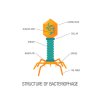
© iStock
Structure of a bacteriophage
In the adult human gut only, the total population of bacteriophages[18] is estimated[19] to 10 e15, which represents between 10 e8 and 10 e10 phages per gram[20] of stool. A paper published in 2021 found that the gut viral population is constituted of more than 142,000 different phage species[21].
Notice that these counts focused solely on bacteriophage viruses, which represents only a portion of the total virus population.
Notice also that this number, although substantial keeps increasing. A similar study conducted in 2020 only identified 33,000 viral species in human gut[22]. This rapid progression, only one year apart, suggests that real number of viral species is way higher than these two counts.
Since the beginning of the 20th Century science keeps discovering new human virus species and the rate of discovery is not weakening[23]:

© Woolhouse
Discovery curve for human virus species.
The discovery of human viruses keeps increasing because the discovery methods are mostly based on the underreported clinical observation:
[T]he process of virus detection and identification is still mostly initiated by clinical observation; […]. However, it is widely believed that the vast majority of human infections are not reported to medical practitioners, and that no etiological agent is formally identified for most of those that are. Thus, it is unsurprising that we are still discovering new viruses that can infect humans[24]
A tiny proportion of viruses are pathogenic, these are the ones who are tracked viruses and sometimes are identified. But most viruses are non–pathogenic and therefore highly likely to stay untracked and un-identified.
A study focused on viral population in the human blood plasma counted a concentration of 10 e7 (10 million) virions[25] per milliliter[26]. During this count, several novel viruses of the anellovirus family were serendipitously discovered[27]
The official number of viruses and the diversity of viral species in our body (guts, blood) are stunningly high and ever-growing. In addition viruses are also markedly present within our own DNA:
One of the most earth-shaking papers of this century was the publication of the human genome sequence. About half, possibly even two-thirds of the sequence are composed of more or less complete endogenous retroviruses (ERVs) and related retroelements (REs) [...].
The origin of REs is being discussed as remnants of ancient retroviral germline infections that became evolutionarily fixed in the genome.
About 450,000 human ERV (HERV) elements constitute about 8% of the human genome consisting of hallmark retroviral elements like the gag, pol, env genes and flanking long terminal repeats (LTR) that act as promoters. Howard Temin, one of the discoverers of the reverse transcriptase, in 1985 already described endogenous retrovirus-like elements, which he estimated to about 10% of the human and mouse genome sequence.
The actual number is about 45% as estimated today. In some genes such as the Protein Kinase Inhibitor B (PKIB) gene we determined about 70% retrovirus-related sequences. Is there a limit? Could it have been 100%?" [28]
The prevalence of virus-related sequences (LINE, SINE, ERV and transposons) in the human genome is illustrated by the chart below:

© Weiss
Proportion of transposable elements in the human genome
The human genome is not the only one revealing a large proportion of ERVs. Viral sequences also constitute about half of the mammalian genome:
For example, ERV sequences encompass 42.2% of the human genome and almost half of the mammalian genome[29]
In total, the human genome contains nearly half a million viral sequence insertions[30] along with about 200,000 copies of ERVs, which have been introduced through at least 31[31] infection events[32]. Notice that these counts only reflect the number of identified viral sequences and retrovirus.
Our DNA code is literally made of viral sequences. This omnipresence is compounded by the numerous copies of the same viral sequences found all over the human genome:
[...] humans, given the vast proliferation of insertion sites involving those 30 to 50 families of viruses, and the 200 or so subgroups. If, for example, we look at just the HERV clone 4-1 variant of the HERV-E family that was studied by Sekigawa in relation to SLE, the human genome contained 85 copies of the virus at various integration sites within different chromosomes. [33]
In a world which is so overrun by viruses, and given the proliferation of life on this planet, it is thus evident that viruses sicken and kill only rarely and their purpose goes far beyond the thin visible veneer of viral diseases.
Evidence proving that viruses are much more than pathogenic agents is the fact that viruses evolve, on average, about one million times faster than their hosts[34]. How could hosts develop any effective defense system against such an ever-changing “enemy”?
[1] Hendrix, Roger W. et al. (1999) “Evolutionary relationships among diverse bacteriophages and prophages: All the world’s a phage” PNAS 96 (5) 2192-2197
[2] K. Moelling (2020) “Viruses More Friends than Foes” Electroanalysis 32, 669
[3] Wu, Katherine J. (2020) "There are more viruses than stars in the universe. Why do only some infect us? - More than a quadrillion quadrillion individual viruses exist on Earth, but most are not poised to hop into humans. Can we find the ones that are?" National Geographic Society
[4] Mackie, Glen (2002) "To see the Universe in a Grain of Taranaki Sand". Swinburne University of Technology
[5] 6X10⁻⁵ cubic inch
[6] Emiliani, C. (1995). “Evolution--a composite model“. Evolutionary Theory Vol.10, No.6, 299-303
[7] 1012 to 1014 viruses per cubic inches.
[8] One drop equals 50 cubic millimeters
[9] The weight of one virus is roughly equal to 10-18g. See:
Bahr GF et al. (1976) “Determination of the mass of viruses by quantitative electron microscopy”. Q Rev Biophys.;9(4):459-89
[10] Foulsham, George (2011) “10 million viruses in one drop of seawater” UC Santa Barbara
[11] Racaniello, Vincent (2013) “How many viruses on Earth?” Virology Blog
[12] Ibid
[13] Archaea are single-celled microorganisms with structure similar to bacteria. They are evolutionarily distinct from bacteria and eukaryotes and form the third domain of life. See:
Suchodolski, Jan. (2013) “Canine and Feline Gastroenterology”, Chapter 2 - Gastrointestinal Microbiota, Pages 32-41. Ed. Robert J. Washabau and Michael J. Day
[14] Joseph, Rhawn (2009) “The Evolution Of Life From Other Planets Part 1”. Journal of Cosmology. Vol 1, 100-150
[15] Le Libre Editors (2021) “Combien de virus inhale-t-on chaque minute?’’ Le Libre
[16] Łusiak-Szelachowska, M. et al. (2020) “The Presence of Bacteriophages in the Human Body: Good, Bad or Neutral?” Microorganisms, 8(12), 2012
[17] Bianconi E, et al. (2013) “An estimation of the number of cells in the human body”. Ann Hum Biol. 40(6):463-71
[18] The estimated population of phages in the gut and in the human body is roughly the same because most phages are located within the gut.
[19] Łusiak-Szelachowska, M. et al. (2020) ‘’The Presence of Bacteriophages in the Human Body: Good, Bad or Neutral ?’’ Microorganisms, 8(12), 2012
[20] Babickova, J et al. (2015) “Pathological and Therapeutic Interactions between Bacteriophages, Microbes and the Host in Inflammatory Bowel Disease” World J. Gastroenterol. 21, 11321–11330
[21] Camarillo-Guerror, L. F. et al. (2021) “Massive expansion of human gut bacteriophage diversity”. Cell 184, 1098–1109.E9
[22] Gregory AC et al. (2020) “The Gut Virome Database Reveals Age-Dependent Patterns of Virome Diversity in the Human Gut”. Cell Host Microbe. 28(5):724-740.e8
[23] Woolhouse, Mark et al. (2008) “Temporal trends in the discovery of human viruses”. Proceedings. Biological sciences / The Royal Society. 275. 2111-5. 10
[24] Woolhouse MEJ, Adair K. (2013) “The diversity of human RNA viruses” Future Virol. 8(2):159-171
[25] A virion is an entire virus particle, consisting of an outer protein shell called a “capsid” and an inner core of RNA or DNA
[26] 0.035 fl
[27] Mya Breitbart, Forest Rohwer (2005) “Method for discovering novel DNA viruses in blood using viral particle selection and shotgun sequencing” BioTechniques 39:5, 729-736
[28] Moelling, K. & Broecker, F. (2019) “Viruses and Evolution - Viruses First? A Personal Perspective” Frontiers in microbiology, 10, 523
[29] Wickramasinghe, Chandra et al. (2013) “Diseases From Space: Astrobiology, Viruses, Microbiology, Meteors, Comets, Evolution” Cosmology Science Publishers
[30] Feschotte, C., Gilbert, C. (2012) “Endogenous viruses: insights into viral evolution and impact on host biology” Nature Review Genetics 13, 283–296
[31] This figure is reminiscent of the estimated 20 to 30 extinction events that our planet experienced. See chapter “Mass extinctions”
[32] Rhawn, Joseph (2010) “Comets and Contagion: Evolution, Plague, and Diseases From Space” Journal of Cosmology
[33] Ryan, 2013
[34] Wain-Hobson, S. (2008) “Retrovirus evolution”. In: Origin and Evolution of Viruses. Academic Press